
Fluorescence Microscopy
Background
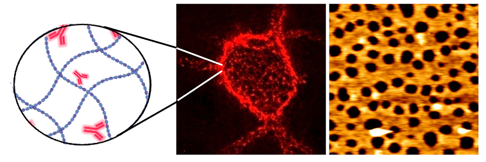
Fluorescence microscopy is a specialised imaging technique, which enables visualisation of fluorescently tagged molecules. Fluorescent tags are added to antibodies which bind specifically to cellular components, therefore labelling the specific component which can be imaged using a fluorescence microscope.
For example, the glycosaminoglycan hyaluronan, abundant in extracellular matrices can be tagged by antibodies conjugated to a fluorophore to fluorescently label hyaluronan and analyse its presence in different extracellular environments. Richter Labs have found the hyaluronan matrix surrounding mammalian oocytes during ovulation and fertilization to be the softest elastic biological material reported to date. We reconstitute hyaluronan matrices in vitro to study the biochemical and physical mechanisms underpinning their self-organisation, and how their unique properties guide cell behaviour. This provides important new insight as to how the complexity of glycan-protein interactions contributes to physiological processes as diverse as inflammation and ovulation, immune cell recruitment, neuronal plasticity and cartilage function. Read more about it here.
.png)
Applications in our research
To learn more about the research involving fluorescence microscopy within Richter Labs, please click the icons below:
